
The human body contains approximately 650 muscles and the different sets of muscles are enveloped by a membrane of connective tissue called the fascia (e.g., thoracolumbar fascia).
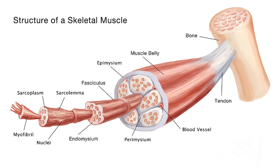
The functional and structural unit of muscle tissue is the muscle fiber, differentiated muscle cell or myocyte. Each muscle fiber is surrounded by a fine network of reticular fibers (endomysium). In turn, the muscle fibers are grouped in bundles, among which we find connective tissue structures (perimysium) with collagen and elastic fibers, vessels and nerves. The entire muscle is in turn covered by a connective tissue sheath (epimysium), which continues with the connective tissue that surrounds the fiber bundles and tendons.

The cell membrane is called a sarcolemma. The cytoplasm is called sarcoplasm. In it, we find the intracellular fluid (hyaloplasm or cytosol) and a set of cellular organelles responsible for carrying out the functions and nutrition of the muscle cell.
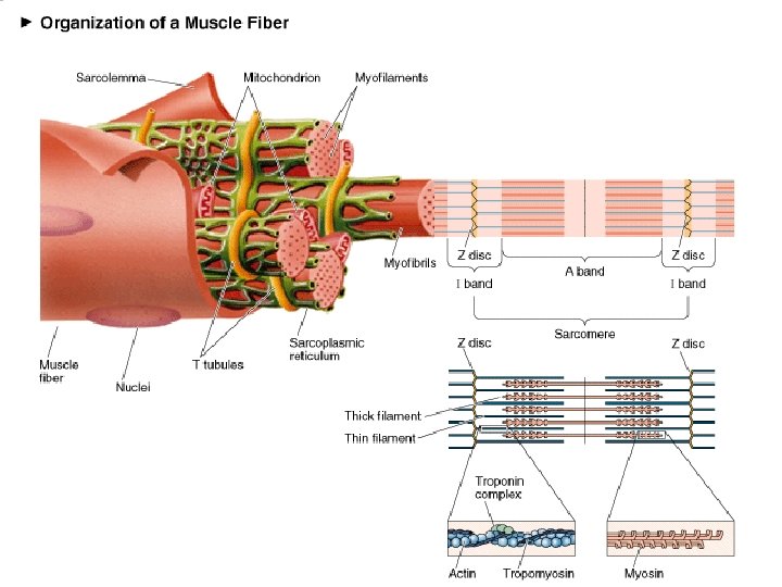
Muscle fibers contain little cytosol, and the vast majority of the cytoplasm is occupied by complex structures called myofibrils, which are bundles of elastic and contractile proteins that carry out the function of muscle contraction.
The muscle fibers contain an extensive sarcoplasmic reticulum, which is arranged around the myofibrils. The function of the sarcoplasmic reticulum is to concentrate and sequester calcium ions (Ca2 +). In close association with the sarcoplasmic reticulum, we find the T tubules or transverse tubules, invaginations of the sarcolemma that penetrate into the fiber perpendicular to the surface, and inside we find extracellular fluid. The T tubules allow the action potential that originates from the cell surface in the motor endplate to spread until it reaches the interior of the muscle fiber.
Each muscle fiber contains more than 1,000 myofibrils, which are composed of proteins of various types:
- Contractile proteins: actin and myosin
- Modulator proteins: tropomyosin and troponin
- Accessory giant proteins, which give the muscle elasticity: titin and nebulin
Myosin is the fibrous protein constituent of thick filaments. It is the most abundant protein in skeletal muscle.
Actin is a globular protein that can be found in the form of a monomer (actin G) or forming fine filaments (actin F).
Tropomyosin and troponin are found in the fine filaments associated with actin. They prevent cross-bridges (the junction between actin and myosin) from forming during relaxation and triggering muscle contraction.
- Tropomyosin covers the binding sites of actin with myosin when the muscle is in a resting state.
- Troponin binds to calcium during muscle contraction causing a change in the molecule that allows tropomyosin to be displaced, exposing the actin-myosin binding sites.
Titin is a huge elastic molecule that stabilizes the position of the contractile elements and recovers the length of the muscles during relaxation or the resting position. Contributes to the generation of passive muscle tension. It is the largest known protein.
Nebulin is a non-elastic giant protein that contributes to the alignment of fine filaments.
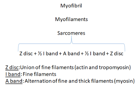
Among all these proteins that make up a myofibril, junctions and gaps (bands, lines and areas) are defined that delimit the structure of the functional unit of a myofibril: the sarcomere.
Bibliographic references:
- Marchante D. (2020). Physiology of exercise and muscle hypertrophy. Basic concepts [PDF file]. Recoverde from https://universidadpowerexplosive.com/

Leave a comment